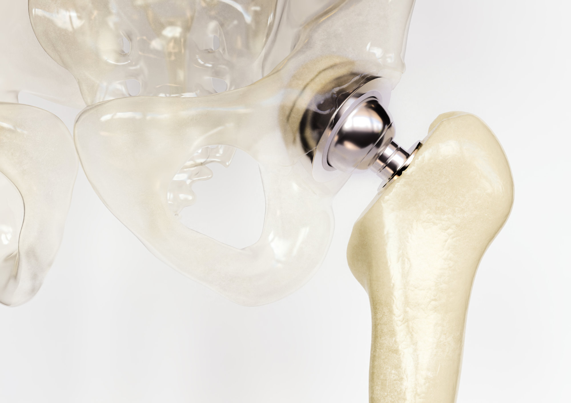
Anatomisch korrekte 3D-Visualisierung — Hüftgelenk mit Arthrose-Verschleiß und das künstliche Gelenkimplantat. Für Patientenaufklärung und medizinische Fachkommunikation. Anatomically correct 3D visualisation — hip joint with osteoarthritis wear and the artificial joint implant. For patient education and medical communication.
Medical-Visualisierung Medical visualisation
Anatomisches Modeling, Lighting, Rendering Anatomical modeling, lighting, rendering
Cinema 4D · Redshift
Medizin · Implantat-Visualisierung Medical · Implant visualisation
Detaillierte 3D-Visualisierung eines Hüftgelenks — einmal mit fortgeschrittener Arthrose (Knorpelverschleiß am Hüftkopf), einmal mit dem künstlichen Gelenkimplantat. Beide Darstellungen basieren auf medizinischen Referenzdaten und zeigen Knochenstruktur, Knorpel und Implantatkomponenten so, dass sie sofort visuell unterscheidbar sind.
Anatomisch korrekte Medical-Visualisierung — insbesondere die Darstellung von Implantaten — ist mein Spezialgebiet. Die Bilder werden in der Patientenaufklärung, in Fachpublikationen und bei medizinischen Präsentationen eingesetzt: komplexe orthopädische Sachverhalte werden so verständlich und anschaulich.
Detailed 3D visualisation of a hip joint — one with advanced osteoarthritis (cartilage wear on the femoral head), one with the artificial joint implant. Both renderings are based on medical reference data and present bone structure, cartilage and implant components so they are immediately visually distinguishable.
Anatomically correct medical visualisation — particularly implant visualisation — is my specialty. The images serve in patient education, in professional publications and at medical presentations: they make complex orthopaedic matters understandable.
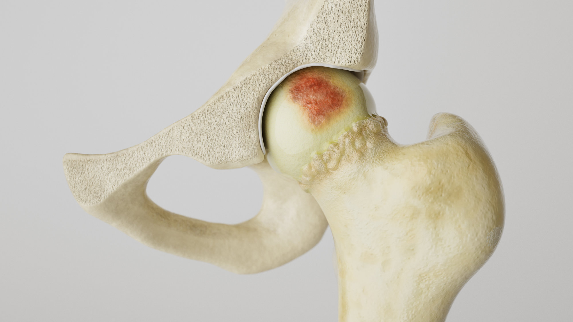
Links: Hüftgelenk mit Arthrose-Verschleiß. Rechts: Künstliches Hüftgelenk-Implantat. Left: Hip joint with osteoarthritis. Right: Artificial hip joint implant.