Project
Medical stock image · Osteoporosis
Services
Anatomical modeling, lighting, rendering
Software
Cinema 4D · Redshift
Field
Patient education · Professional publications
About the project
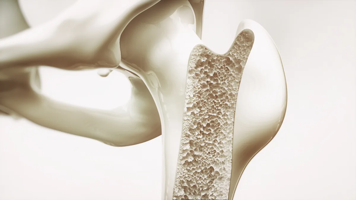
A two-part visualisation of osteoporosis. The first image shows the progression of bone-density loss in the femoral neck across four stages — from the dense, healthy spongiosa on the left to the depleted trabecular structure of advanced osteoporosis on the right. The sequence is particularly suited to patient education, making the disease progression understandable at a glance.
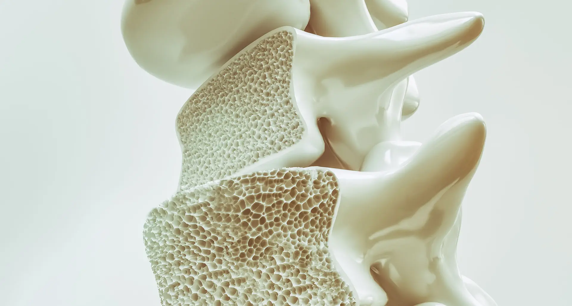
In the second image, the cross-sectioned vertebral body reveals the inner spongiosa in detail — the characteristic honeycomb-like structure of trabecular bone, whose thinning increases fracture risk. Both visualisations were modeled with anatomical accuracy and rendered in a quiet studio lighting that emphasises the material quality of bone without distracting from the medical message.
Anatomically correct medical visualisation is my specialty. The images serve in patient education, in medical advice portals and in professional publications, making complex medical content understandable.
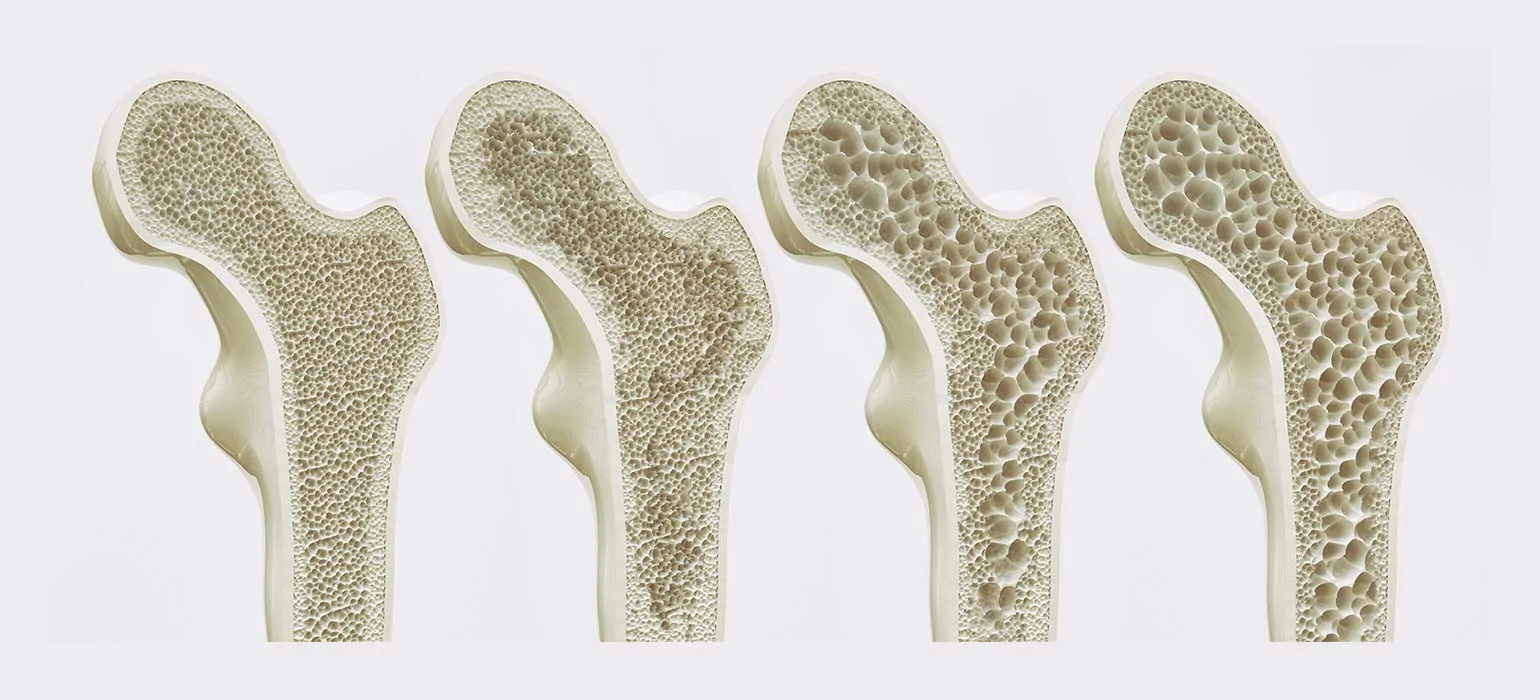
Femoral neck in longitudinal section — four stages of bone-density loss, from healthy to advanced osteoporosis.
Vertebral body cross-section — detail of the trabecular spongy bone structure.
Publications
These visualisations are licensed as stock imagery for medical online portals and specialist publications. A selection of where they appear:
pflege.de — the stages of bone-density loss in the femoral neck illustrate the osteoporosis health guide.
View publication →
Deutsches Ärzteblatt — the cross-sectioned vertebral body with its trabecular spongy structure accompanies the specialist articles on osteoporosis.
View publication →
dieapotheke.com — a close-up of advanced bone-density loss illustrates the osteoporosis guide in the pharmacy blog.
View publication →